Changes in brain activity elicited with low-intensity focused ultrasound
Research by Vincent Koppelmans, Tom Riis, Rana Jawish, Eric Garland, Taylor Webb, Jan Kubanek, and Brian Mickey
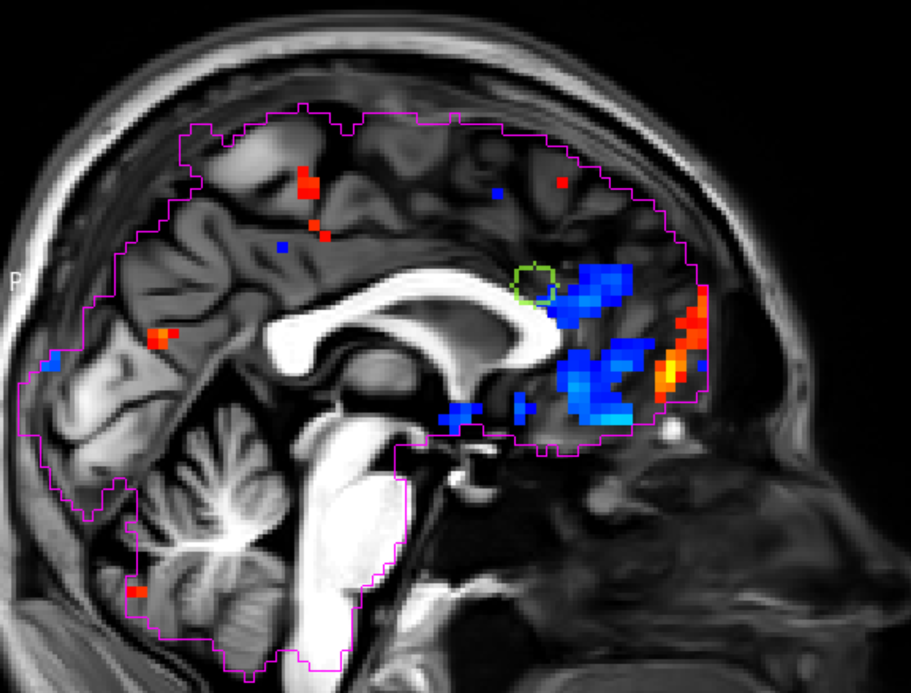
The image below represents changes in brain activity elicited with low-intensity focused ultrasound. This individual was using opioid medication for chronic pain. The front of the head is to the right. The green box indicates the location where ultrasonic stimulation was delivered during functional MRI. Red–yellow colors indicate brain activation and blue colors represent deactivation. This project relies on CHPC resources.
Attribution: This content was provided by researchers involved with the project and edited by staff at the CHPC.